
Global organ shortage results in thousands of patients dying annually while on transplant waitlists, with lungs among scarcest organs. Lung transplantation is limited by donor availability, ischemia time of only a few hours, and strict compatibility criteria. Chronic lung diseases such as COPD, pulmonary fibrosis, and lung cancer represent leading causes of morbidity and mortality worldwide, driving demand for advanced regenerative solutions.
3D bioprinting using patient-derived cells offers potential to reduce rejection and long-term immunosuppression needs. Regenerative strategies using partial lung repair or segmental tissue replacement could decrease the need for full-organ transplants in selected indications.
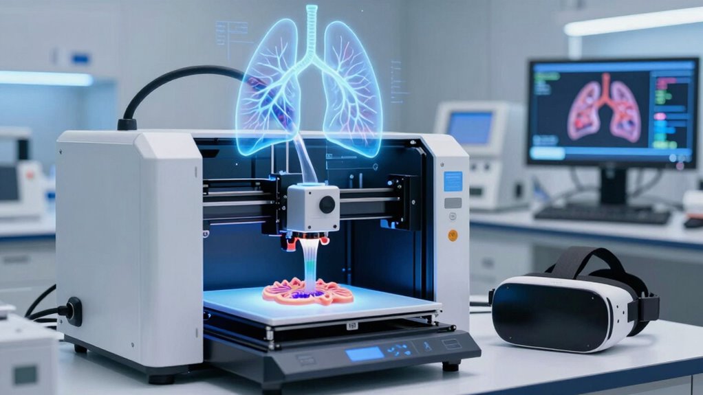
3D bioprinting is defined as layer-by-layer deposition of bioink containing cells and biomaterials to create functional, living 3D structures. The process typically involves pre-bioprinting imaging and modeling, printing with bioink, and post-printing maturation and functional testing. Key requirements for lung constructs include replication of branching airways, alveolar sacs, and integrated vascular networks for gas exchange.
Bioinks combine live cells with biocompatible hydrogels or polymers, tuned for mechanical compliance similar to native lung parenchyma. Scaffold design must support air flow, elasticity, and vascular perfusion while maintaining structural stability under respiratory cycles.
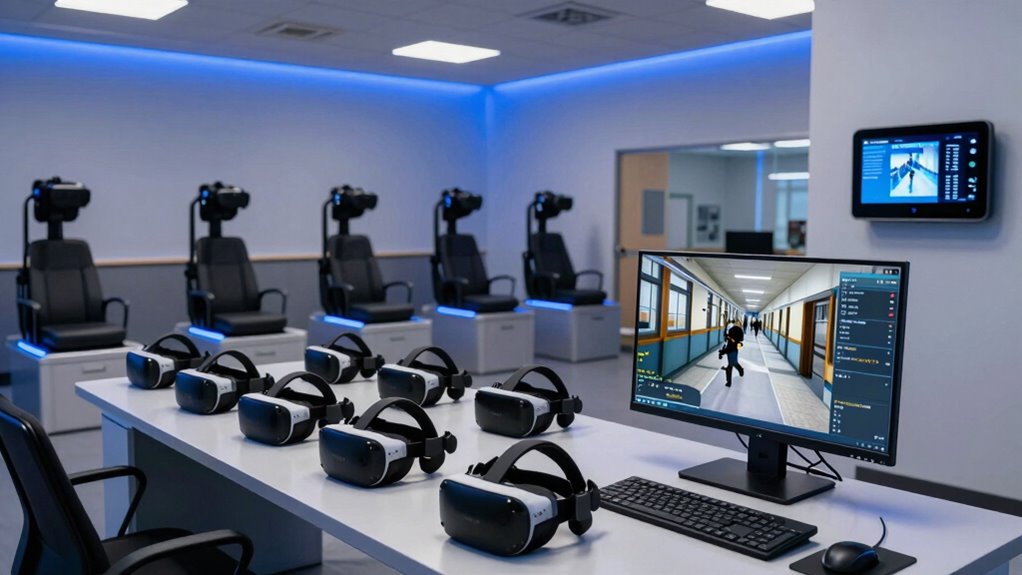
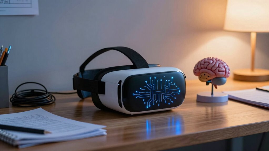
McGill University Health Centre (MUHC) teams employ VR headsets to visualize high-resolution digital lung models for pre-print design. VR systems allow interactive exploration of complex bronchial trees and vascular structures to identify ideal repair or replacement zones. Patient-specific CT or MRI data are imported into VR platforms, enabling customized 3D modeling of airways and lesions.
VR-guided planning supports the definition of exact geometry for 3D-printed lung patches or bridges to match local anatomy. Combined VR and 3D printing workflows aim to improve the precision of segmental reconstruction and minimize resection of healthy tissue.
MUHC-associated researchers in Montreal are developing 3D-printed lung “patches” or “bridges” to bypass damaged segments and reconnect healthy tissue. Printed constructs use lung-specific biomaterials designed to be biocompatible and support ingrowth of new blood vessels from host tissue. These technologies could become standard medical applications as specialized robots for bioprinting become common in hospitals by 2030.
Preclinical tests in mice show no significant rejection signal, indicating promising local biocompatibility of the printed materials. Patches are intended for localized reconstruction in conditions such as airway or lung cancers where only part of the lung is compromised.
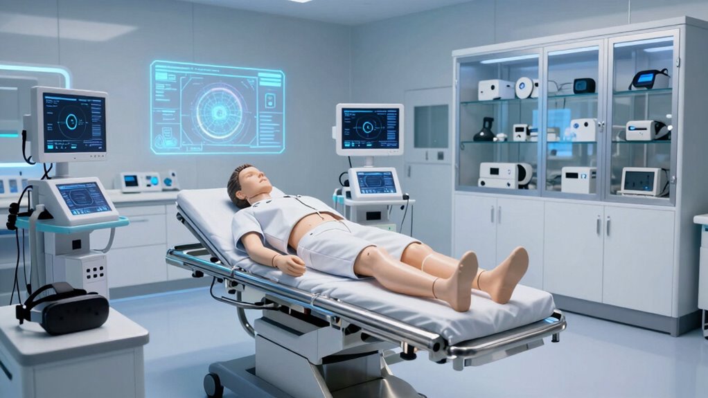
This strategy is designed to preserve functioning lung parenchyma, potentially avoiding or delaying full-organ transplant dynamic, real-time intraoperative guides. The technology also enhances surgical team capabilities through advanced technology training and collaboration.
References
- https://accscience.com/journal/IJB/11/6/10.36922/IJB025350357
- https://www.youtube.com/watch?v=4-PCM8s6nLM
- https://pmc.ncbi.nlm.nih.gov/articles/PMC8252697/
- https://www.regmednet.com/virtual-reality-and-3d-printing-prepares-anesthesiologists-for-pediatric-lung-surgery/
- https://knowhow.distrelec.com/3d-printing/how-3d-bioprinting-could-save-millions/
- https://www.youtube.com/watch?v=G6Jw5-TgA7k
- https://3dprintingindustry.com/news/muhc-researchers-test-vr-and-3d-printing-for-better-lung-repair-247926/
- https://medicalfuturist.com/3d-bioprinting-overview